Common Eye Conditions
Understanding Your Eye
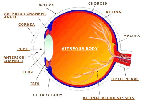
The structure and functioning of the eye are, in many ways, similar to that of an autofocus camera.
The Cornea
At the front of the eye is the cornea. This is the clear transparent curved surface that allows light rays and images into the eye. It also plays a significant role, together with the lens, in focusing these images onto the retina. Hence, it is this corneal curvature that LASIK seeks to modify to correct for refractive errors in the eye. The clarity of the cornea is also affected by disease, infection and degenerations and in severe cases, corneal transplant surgery may be needed to restore clarity to the cornea and improve eyesight.
The Anterior Chamber, Iris and Pupil
Just behind the cornea is the anterior chamber, a space filled with a clear liquid known as the aqueous. This is produced by the ciliary body and its function is to provide nutrients to the cornea and the lens. The iris is the coloured part of the eye with an aperture (or hole) in the middle called the pupil. It controls the size/diameter of the pupil thereby adjusting the amount of light that enters the eye. This is similar to the function of the diaphragm of a camera controlling the aperture size.
Inflammation of this iris/ciliary body structure causes a condition called Iritis or Uveitis and this can be the result of various local or systemic diseases.
The Anterior Chamber Angle
The anterior chamber angle is the area where the iris is attached to the peripheral corneal limbal region. It has the tissues (trabecular meshwork) that are responsible for the reabsorption of the aqueous from the anterior chamber. Abnormalities in this area can cause the condition called Glaucoma. If the trabecular meshwork is physically blocked by the root of the iris, Closed Angle Glaucoma results. If it is the malfunction of the trabecular meshwork itself that results in poor reabsorption of aqueous in the presence of an open angle, then Open Angle Glaucoma can occur.
The Lens
The clear lens plays a major role together with the cornea in focusing images onto the retina. It is held in place by suspensory ligaments which are attached to the ciliary body. Contractions of the ciliary body exert forces onto the suspensory ligaments which will in turn affects the curvature of the lens to adjust accordingly and focus the images, whether near or far, onto the retina. As one ages, the clarity of the lens starts to change and cloudiness of the lens results in Cataract formation. If vision is significantly affected, the removal of the lens surgically and replacing it with an artificial intraocular lens implant (IOL) will help restore sight.
Vitreous Body
The vitreous is a transparent gel like substance that fills the globe of the eye. As one ages, the gel like nature of the vitreous becomes more liquid and it is at this stage that precipitates can sometimes form within the vitreous causing one to see Floaters. The liquification and contraction of the vitreous can also cause one to see flashes and in some cases cause retinal holes, tears or detachment.
The Retina
The retina is the nerve tissue of the eye that contains photoreceptors that respond to light and images (this is equivalent to the film in a camera.) These responses are converted to electrical impulses and transmitted to the brain via the optic nerve. There are 2 types of photoreceptors; Rods, which are more widely distributed in the retina, are responsible for peripheral and night vision, and Cones, which are mostly concentrated in and around the central portion of the retina called the macula. The cones are mainly responsible for sharp central vision and our appreciation of colour. It is here that a condition known as Age Related Macular Degeneration can affect the central vision of a person as he grows old.
Tears and holes in the retina can also result in Retinal Detachments which if not treated urgently can result in permanent vision loss.
The Retinal Blood Vessels
The retinal blood vessels are fine vessels comprising both arteries and veins. They carry blood and provide oxygen and nourishment to the retina. Diseases like diabetes and hypertension can affect these blood vessels, resulting in a group of conditions known as Retinal Vascular Disorders.
The Optic Nerve
The optic nerve transmits the electrical impulses generated by the retina, in response to images, to the brain. In glaucoma, the optic nerve head may be compromised due to the increased intraocular pressure and ‘cupping’ of the optic nerve results. This may lead to loss of peripheral vision and if left untreated, the visual loss will progress until central vision is also affected. Inflammations of the optic nerve can also occur (optic neuritis) leading to loss of vision.
Common Eye Conditions
- Age Related Macular Degeneration
- Blepharitis
- Cataracts
- Central Serous Chorioretinopathy
- Contact Lens Problems
- Diabetes and the Eye
- Dry Eyes
- Floaters and Flashes
- Glaucoma
- LASIK Surgery
- Pterygium and Pingueculae
- Refractive Errors
- Retinal Detachment
- Retinal Vascular Disorders
- Uveitis and Ocular Inflammations
- Vision Problems in Children